This manual, like those from Cold Spring Harbor Laboratory, aids learning through practical biology experiments. It supports products like Visible Biology, offering detailed guidance for effective lab work.
Purpose and Scope
This laboratory manual serves as a comprehensive guide for students undertaking practical biology coursework. Its primary purpose is to facilitate a deeper understanding of core biological principles through hands-on experimentation. The scope encompasses a wide range of techniques, from fundamental microscopy and cell biology to advanced molecular genetics and ecological investigations.
Aligned with resources like NCERT guidelines and Visible Biology, it aims to develop essential laboratory skills – observation, data collection, analysis, and interpretation. The manual supports cloning and sequencing explorations, mirroring the detailed protocols found in specialized texts. Ultimately, it prepares students for success in further biology studies and research.
Safety Regulations in the Biology Lab
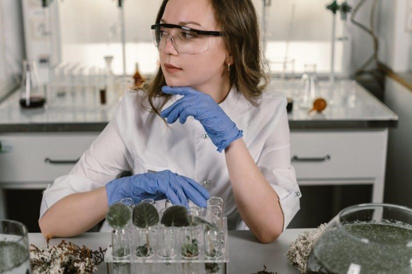
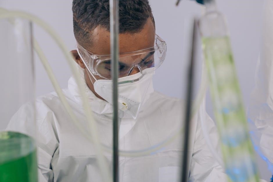
Prioritizing safety is paramount within the biology laboratory. This manual emphasizes strict adherence to established safety protocols to protect students and prevent accidents. Regulations include mandatory eye protection, appropriate attire – lab coats and closed-toe shoes – and proper handling of biological specimens and chemical reagents.
Students must understand disposal procedures for biohazardous waste and be aware of emergency protocols, including the location of safety equipment like eyewash stations and fire extinguishers. Responsible conduct, including no food or drink consumption, is crucial. Following these regulations ensures a secure and productive learning environment for all biology students.
Essential Laboratory Equipment
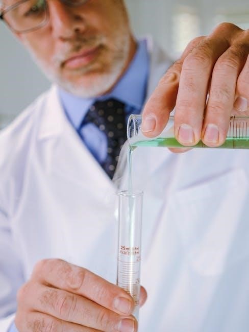
A well-equipped biology lab is crucial for successful experimentation. This manual details the function and proper use of essential equipment, including microscopes – both light and potentially electron – for cellular observation. Other key tools include glassware (beakers, flasks, pipettes), centrifuges for separating substances, and spectrophotometers for analyzing light absorption.
Furthermore, understanding the operation of autoclaves for sterilization, and equipment for molecular biology techniques like gel electrophoresis is vital. Proper maintenance and careful handling of all equipment are emphasized to ensure accurate results and longevity of the lab’s resources.
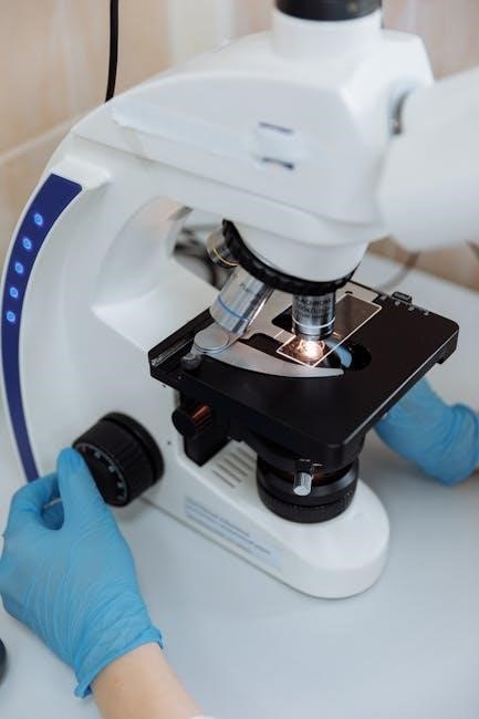
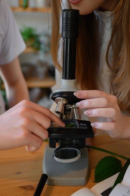
Microscopy Techniques
Microscopy is fundamental to biology, utilizing light and electron microscopes to visualize cells and tissues, requiring careful slide preparation for optimal viewing.
Types of Microscopes (Light, Electron)
Microscopy employs diverse techniques, broadly categorized into light and electron microscopy. Light microscopes, the more accessible option, utilize visible light and lenses to magnify specimens, ideal for observing cells and tissues. These are frequently used with prepared slides, as suggested by lab manuals.
Electron microscopes, however, offer significantly higher magnification and resolution, employing beams of electrons instead of light. This allows visualization of subcellular structures with incredible detail. There are two main types: Transmission Electron Microscopy (TEM) and Scanning Electron Microscopy (SEM), each providing unique insights into biological samples. Understanding these distinctions is crucial for effective biological investigation.
Preparing Slides for Microscopy
Proper slide preparation is paramount for clear microscopic observation. Begin with a clean glass slide and coverslip; For wet mounts, place the specimen in a drop of liquid, gently lower the coverslip to avoid air bubbles, and secure if needed.
Fixed specimens, often requiring staining, necessitate careful mounting procedures. Staining enhances contrast, highlighting cellular structures. Lab manuals emphasize techniques like sectioning, dehydration, and embedding for optimal results. Accurate labeling is essential for identification. Careful preparation minimizes artifacts and ensures reliable data collection during observation.
Observing Cells and Tissues
Microscopic observation reveals intricate cellular details. Begin with low power objectives to locate the specimen, then increase magnification for closer examination. Focus carefully, adjusting both coarse and fine focus knobs.
Identify key structures – nucleus, cytoplasm, cell wall (in plant cells). Observe tissue organization, noting cell arrangement and intercellular spaces. Lab manuals often guide identification of specific cell types. Accurate sketching and detailed notes are crucial for recording observations. Remember proper lighting and contrast enhance visibility, aiding in accurate analysis.
Cell Biology Experiments
Explore cellular processes like structure, function, permeability, and division (mitosis/meiosis). These experiments, detailed in lab manuals, build foundational biology understanding.
Cell Structure and Function Observation
This experiment focuses on visualizing and understanding the fundamental components of cells, the basic units of life. Utilizing microscopy – both light and potentially electron – students will prepare slides to observe various cell types, including plant and animal cells.
Detailed observation will highlight key structures like the nucleus, cytoplasm, cell membrane, and, in plant cells, the cell wall and chloroplasts. The lab manual will guide students in identifying these structures and correlating their morphology with specific cellular functions.
Understanding these relationships is crucial for grasping broader biological concepts. Careful data recording and analysis, as outlined in the appendix, are essential components of this exercise.
Cell Membrane Permeability
This lab investigates the selective permeability of cell membranes, a critical characteristic enabling cellular function. Students will explore how different molecules – based on size and charge – traverse the membrane. Experiments may involve observing osmosis using varying solute concentrations and membrane types.
The manual details procedures for setting up controlled experiments, carefully recording observations, and analyzing data to determine permeability rates. Understanding factors influencing permeability, like temperature and membrane composition, will be emphasized.
Proper data analysis, referencing the appendix, is vital for interpreting results and drawing conclusions about membrane transport mechanisms.
Mitosis and Meiosis Observation
This section guides students through observing the distinct stages of mitosis and meiosis using microscopy. Prepared slides of plant or animal cells undergoing cell division will be utilized, allowing for identification of chromosomes, spindle fibers, and daughter cells.
The manual provides detailed diagrams and descriptions of each phase – prophase, metaphase, anaphase, and telophase – for both processes. Students will learn to differentiate between mitosis, resulting in identical cells, and meiosis, producing genetically diverse gametes.
Accurate observation and recording of cell structures are crucial for understanding these fundamental processes.
Molecular Biology Techniques
This section details methods like DNA extraction, purification, gel electrophoresis, and PCR, mirroring techniques described in manuals from Cold Spring Harbor Laboratory Press.
DNA Extraction and Purification
DNA extraction is a foundational molecular biology technique, isolating genetic material from cells or tissues. This process typically involves cell lysis, using detergents to disrupt membranes, followed by removal of proteins and RNA via enzymatic digestion or chemical methods.
Purification steps, often employing phenol-chloroform extraction or spin columns, ensure high-quality DNA free from contaminants; The resulting DNA is then suitable for downstream applications like PCR, gel electrophoresis, and sequencing. Manuals, such as those referenced from Cold Spring Harbor Laboratory, provide detailed protocols for optimizing these procedures, ensuring reliable and reproducible results. Successful extraction yields DNA ready for further molecular analysis.
Gel Electrophoresis

Gel electrophoresis separates DNA fragments based on size and charge. This technique utilizes an agarose or polyacrylamide gel matrix, through which DNA migrates under an electric field; Smaller fragments move faster and further than larger ones, creating distinct bands visible after staining with ethidium bromide or alternative dyes.
Laboratory manuals, like those from Cold Spring Harbor, detail proper gel preparation, sample loading, and running conditions. Gel electrophoresis is crucial for verifying DNA extraction success, analyzing PCR products, and preparing samples for downstream applications like cloning and sequencing. Accurate interpretation of band patterns is essential for reliable results.
Polymerase Chain Reaction (PCR)
Polymerase Chain Reaction (PCR) is a fundamental molecular biology technique used to amplify specific DNA sequences. This process involves repeated cycles of heating and cooling, allowing DNA polymerase to synthesize new DNA strands complementary to a target sequence. Essential components include DNA template, primers, nucleotides, and a heat-stable DNA polymerase.
Laboratory manuals, such as those referenced, provide detailed protocols for optimizing PCR conditions, including primer design, annealing temperature, and cycle number. PCR is widely used in cloning, sequencing, and diagnostic applications, enabling researchers to generate millions of copies of a desired DNA fragment from a minimal starting amount.

Genetic Experiments
This section details cloning and sequencing explorations, often spanning 68 weeks, focusing on analyzing genes like GAPC, as detailed in laboratory manuals.
Mendelian Genetics and Punnett Squares
This lab component focuses on foundational principles of inheritance, utilizing Punnett squares to predict genetic outcomes. Students will explore monohybrid and dihybrid crosses, analyzing phenotypic and genotypic ratios derived from experimental data.
Practical exercises will involve observing traits in simulated organisms or simple biological systems, allowing for direct application of Mendelian laws. Emphasis is placed on understanding concepts like dominant and recessive alleles, segregation, and independent assortment.
The manual provides detailed instructions for setting up crosses, collecting data, and interpreting results, reinforcing theoretical knowledge with hands-on experience. This builds a strong base for more complex genetic analyses.
DNA Cloning and Sequencing
This section guides students through the process of isolating and replicating specific DNA fragments – DNA cloning. Utilizing techniques described in resources like those from Cold Spring Harbor Laboratory Press, the manual details vector preparation, ligation, and transformation procedures.
Students will learn to analyze cloning results, confirming successful insertion of DNA into vectors; Furthermore, the lab explores DNA sequencing, enabling determination of the nucleotide order within cloned fragments.
The Cloning and Sequencing Explorer series, a 68-week curriculum, provides a comprehensive framework. Emphasis is placed on understanding the principles and applications of these vital molecular biology techniques.
Physiological Experiments
This section investigates biological processes within living organisms, focusing on enzyme activity, photosynthesis, and respiration – key areas of plant and animal physiology;
Enzyme Activity and Factors Affecting It
Enzyme activity is a cornerstone of physiological investigations, demonstrating how these biological catalysts accelerate reactions within living systems. This lab explores quantifying enzyme function, often utilizing spectrophotometric assays to measure reaction rates.
Crucially, experiments detail how variables like temperature, pH, and substrate concentration influence enzyme efficiency. Students will observe denaturation at extreme pH levels and reduced activity at suboptimal temperatures. Investigating enzyme inhibition – competitive and non-competitive – provides insight into regulatory mechanisms.
Data analysis involves plotting reaction rates against varying conditions, allowing for determination of optimal parameters and Michaelis-Menten kinetics. Understanding these factors is vital for comprehending metabolic pathways and biological control.
Plant Physiology: Photosynthesis and Respiration
Plant physiology experiments within this manual focus on the fundamental processes of photosynthesis and respiration, essential for life on Earth. Labs commonly investigate photosynthetic rates using techniques like measuring oxygen production or carbon dioxide uptake in aquatic plants.
Students will analyze the impact of light intensity and wavelength on photosynthetic efficiency, often employing spectrophotometry. Respiration is explored through monitoring carbon dioxide release or oxygen consumption, demonstrating cellular energy production.
Comparative studies between different plant species or leaf tissues reveal adaptations to varying environmental conditions. Data analysis emphasizes understanding the interconnectedness of these processes and their role in plant growth and survival.

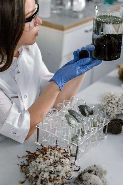
Ecological Investigations
This section details sampling techniques for ecological studies, focusing on biodiversity analysis. Labs guide students in collecting and interpreting data from various ecosystems.
Sampling Techniques in Ecology
Ecological investigations necessitate robust sampling methodologies to accurately represent larger populations and communities. This lab section introduces students to various techniques, including quadrat sampling for plant density, transect methods for environmental gradients, and mark-recapture techniques for mobile animal populations.
Students will learn to design sampling strategies minimizing bias and maximizing statistical power; Emphasis is placed on random sampling, stratified sampling, and systematic sampling, alongside considerations for sample size determination. Proper data collection protocols, including accurate species identification and abundance estimation, are crucial.
Furthermore, the manual details methods for assessing habitat characteristics, such as soil composition, light intensity, and temperature, to correlate with observed species distributions. Understanding these techniques forms the foundation for effective ecological monitoring and conservation efforts.
Analyzing Biodiversity
This section of the laboratory manual focuses on quantifying biodiversity within ecological communities. Students will explore various diversity indices, including species richness, Shannon-Wiener diversity index, and Simpson’s diversity index, to assess community structure.
Practical exercises involve calculating these indices from collected field data, interpreting the results in relation to habitat characteristics, and comparing biodiversity levels across different ecosystems. The manual emphasizes the importance of considering both species richness and evenness when evaluating biodiversity.
Students will also learn to construct species accumulation curves to estimate the completeness of their sampling efforts and assess the potential for discovering new species. Understanding these analytical tools is vital for conservation biology and ecological management.
Appendix
This section provides essential resources: common solution recipes, data analysis guidelines, and a troubleshooting guide for experiments detailed within this biology manual.
Commonly Used Solutions and Reagents
This appendix details frequently utilized solutions crucial for successful biology laboratory work. Recipes include precise concentrations for buffers – Tris, phosphate, and HEPES – essential for maintaining pH stability during experiments.
Detailed instructions are provided for preparing common staining solutions like Gram stain, used for bacterial identification, and methylene blue, valuable for observing cell structures.
Furthermore, this section outlines the preparation of agarose gels for electrophoresis, alongside commonly used loading dyes. Safety precautions regarding reagent handling and proper disposal methods are also emphasized, ensuring a safe and productive laboratory environment for all users of this manual.
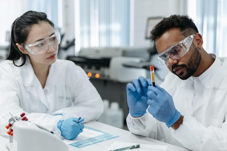
Data Recording and Analysis
Accurate data recording is paramount in any biology laboratory setting. This section emphasizes meticulous note-taking, utilizing pre-formatted tables for organized observation capture; Guidelines detail appropriate units of measurement and significant figure usage for precision.
Furthermore, it provides instruction on basic statistical analysis techniques – calculating means, standard deviations, and performing simple t-tests – to interpret experimental results effectively.
Visual representation of data through graph creation (bar graphs, line graphs) is also covered, alongside proper labeling and axis scaling; Emphasis is placed on objective interpretation and avoiding bias when drawing conclusions from collected data.
Troubleshooting Guide
Experimental setbacks are inevitable in biology. This guide offers solutions to common issues encountered during procedures, from reagent preparation to equipment malfunction. It addresses problems like contamination, unexpected results, and difficulties with microscopy or electrophoresis.
Detailed steps for identifying the source of errors and implementing corrective actions are provided.
The guide also emphasizes safety precautions when dealing with faulty equipment or hazardous materials. It encourages a systematic approach to problem-solving, promoting critical thinking and independent investigation when standard protocols fail to yield expected outcomes, ensuring successful experimentation.